A person holding their knee while running, indicating pain or strain during physical activity.
The knee is the largest and most mechanically complex joint in the body. It bears the equivalent of 3–5 times your body weight during walking, and up to 7–8 times during squatting or climbing stairs. It absorbs impact, provides stability, and enables the full range of lower limb movement. Given what it does, it's perhaps not surprising that knee pain is one of the most common musculoskeletal complaints in India — affecting a huge range of people, from young athletes to elderly adults sitting cross-legged for daily prayers.
Not all knee pain is the same. Where exactly the pain is located, what makes it worse, when it started, and what you were doing at the time all help identify the cause — and the cause determines what helps.
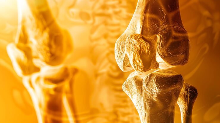
The knee joint involves the femur (thigh bone), tibia (shin bone), and patella (kneecap). Structures that commonly become painful:
Articular cartilage: the smooth surface covering the bone ends. When this wears away, the bones rub together — this is osteoarthritis.
Menisci: two C-shaped discs of fibrocartilage in the knee joint that act as shock absorbers and improve joint congruence. The medial (inner) and lateral (outer) menisci can tear from twisting injuries or degenerate with age.
Ligaments: the ACL, PCL, MCL, and LCL stabilise the knee. Ligament sprains and tears are common in sports and falls.
Patellofemoral joint: the joint between the kneecap and the femur. A very common source of pain, particularly in young people and those who spend a lot of time on stairs or squatting.
Tendons: the patellar tendon (below the kneecap) and quadriceps tendon (above) can become inflamed (tendinopathy) from overuse.
Bursae: fluid-filled sacs around the knee that reduce friction. They can become inflamed (bursitis) from direct pressure or repetitive movement.
Front of the knee (anterior):
Inner side of the knee (medial):
Outer side of the knee (lateral):
Behind the knee (posterior):
Diffuse / throughout the knee:
Cross-legged sitting (padmasana or sukhasana for prayers): sustained deep knee flexion compresses the medial compartment. People who sit cross-legged for hours daily — particularly women — frequently develop medial knee compartment osteoarthritis earlier.
Squatting at Indian toilets: repeated deep knee flexion — particularly with load (standing up from a squat while overweight). This is the joint position of maximum knee joint loading.
Climbing stairs in high-rise apartments: patellofemoral loading. Several times daily, this adds up significantly.
Excess body weight: each additional kilogram of body weight adds approximately 3–5 kg of force across the knee joint during walking. Being 10 kg overweight places an additional 30–50 kg of cumulative load on the knee per step.
Sudden increase in walking or standing: a common trigger — someone who normally sits all day suddenly walks 10,000 steps on a pilgrimage or tour, and their knees aren't ready for the sudden load increase.
Rest from the provocative activity: this doesn't mean complete rest. It means reducing the specific activities that provoke the pain — fewer stairs, less squatting, modified walking distances — while maintaining overall movement.
Ice: for acute flares or after activity, applying ice (wrapped in a towel) for 15–20 minutes reduces inflammation and pain. Particularly useful in the first 48–72 hours after an acute injury or flare.
Compression: a compression bandage or knee sleeve reduces swelling and provides some proprioceptive support.
Elevation: elevating the leg reduces swelling in acutely swollen knees.
Paracetamol and NSAIDs: paracetamol for mild pain. Ibuprofen or naproxen for inflammatory knee pain. Topical diclofenac gel applied to the knee is effective with fewer systemic side effects than oral NSAIDs.
Weight loss: the most effective long-term intervention for osteoarthritis-related knee pain. Even a 5–10% reduction in body weight produces disproportionately large reductions in knee pain because the force reduction applies to every step taken.
The principle is the same as for lower back pain — appropriate movement promotes recovery better than rest.
Straight leg raise:
Lie on your back. One knee bent, the other straight. Tighten the quadriceps of the straight leg (lock the knee) and lift the whole leg to the height of the bent knee. Hold 5 seconds. Lower slowly. 3 sets of 15. This strengthens the quadriceps without loading the knee joint.
Seated knee extension:
Sitting in a chair, slowly extend the knee to fully straight, hold 5 seconds, lower slowly. 3 sets of 15. Builds quadriceps endurance.
Mini squats (shallow):
Stand holding a support. Bend knees to only 20–30 degrees (not a full deep squat). Hold 5 seconds. Return to standing. Builds quadriceps and glutes safely within a pain-free range. 3 sets of 12.
Clamshells:
Lie on your side with knees bent and feet stacked. Without letting the pelvis roll, lift the top knee upward like a clamshell opening. Hold 3 seconds. Lower slowly. 3 sets of 15 each side. Builds hip abductor strength — weak hip muscles are a very common contributor to patellofemoral and general knee pain.
Heel raises:
Stand with feet hip-width apart, holding a support if needed. Rise onto the balls of your feet. Hold 3 seconds. Lower slowly. 3 sets of 15. Builds calf strength which supports knee mechanics.
Step-ups (short step):
Using a low step (10–15 cm), step up and down one foot at a time. 3 sets of 12. Controlled, functional movement that builds knee stability.
Swimming: the best exercise for knee pain — full lower limb movement with essentially no knee joint loading. Backstroke and freestyle are both appropriate. Deep-water walking is excellent.

Man swimming for exercise.
Cycling (low resistance): another excellent low-impact knee exercise. Stationary bike with low resistance and seat height adjusted so the knee reaches only slight flexion at the bottom of the pedal stroke.
See a doctor if:
Investigation: X-ray (for bone and joint space), MRI (for soft tissue — meniscus, ligaments, cartilage). Blood tests if inflammatory arthritis or infection is suspected.
At Prakash Hospital Noida, our orthopaedic team and physiotherapists evaluate knee pain — from sports injuries to osteoarthritis — with clinical assessment, X-ray and MRI where indicated, injection therapy for appropriate cases, physiotherapy referral, and orthopaedic surgical consultation for cases requiring it.
Whether you live in Sector 18, Sector 62, Greater Noida West, or anywhere in the NCR, Prakash Hospital Noida is a trusted name for knee pain, orthopaedics, and sports medicine in Noida.
 Copy
CopyWe offer expert care across key specialties, including Medicine, Cardiology, Orthopaedics, ENT, Gynaecology, and more—delivering trusted treatment under one roof.


Book a consultation with Prakash Hospital's specialists — 24/7 emergency care, 100+ doctors, NABH accredited.

Prakash Hospital Pvt. Ltd. is a 100 bedded NABH NABL accredited multispecialty hospital along with a center of trauma and orthopedics. We are in the service of society since 2001.
OUR SPECIALITIES
Patient Services
PROCEDURES
Contact Us
D – 12A, 12B, Sector-33, G. B. Nagar, Noida, Uttar Pradesh 201301
+91-8826000033


© 2026 All rights reserved.
Designed and Developed by Zarle Infotech