


The Link Between Stress and Heart Disease
Learn symptoms, risk factors, prevention tips and when to seek expert cardiac care in Noida.
25 Mar 2026
by Prakash Hospital
Thin tubes measure pressure and oxygen inside your heart and lung arteries directly, when scans can't settle a question about your valves or the pressure in your lungs.

Sometimes the tests don't agree. The echo calls your valve narrowing moderate, yet you're breathless walking to the gate. Or a scan suggests high pressure in your lungs, and before you start lifelong medicines your doctors need to know exactly how high it is, and why.
Cardiac catheterisation measures from inside the heart. A cardiologist passes a thin tube (catheter) through a vein or artery into the heart chambers and the lung artery, records the pressure in each, takes small blood samples for oxygen and calculates how much blood the heart pumps each minute. A right heart study goes in through a vein and covers the right side of the heart and the lungs. A left heart study goes through an artery to the main pumping chamber and the aortic valve.
It's related to coronary angiography but asks a different question. An angiogram takes X-ray pictures of the heart's own arteries to find blockages, while catheterisation measures how the heart and lungs are working. The two are often combined, for example before valve surgery.
Cardiac catheterisation is one of the diagnostic tests offered by our cardiology department, alongside ECG, 2D echo with Doppler, TMT and Holter monitoring.
Catheterisation usually follows an echo or other scans, when the answer affects a major decision such as surgery, a valve procedure or long-term medicines.
A balloon-tipped catheter enters a vein in the neck, arm or groin and floats through the right side of the heart into the lung artery. It records pressure at each point, including a "wedge" pressure that reflects the left side of the heart, and measures cardiac output, usually without any dye.
A catheter passes from the wrist or groin artery across the aortic valve into the left ventricle, measuring the pressure there and the pressure drop across the valve. A coronary angiogram is usually done at the same time.
In pulmonary hypertension, a medicine may be given to see whether the lung arteries relax, which helps choose treatment. Oxygen samples from several chambers can find and size a hole in the heart.
You lie in the cath lab with ECG stickers and a drip, awake but often mildly sedated. The skin over the vein, and the artery if a left heart study is planned, is numbed, and a short sheath goes into each vessel.
The catheter floats through the right side of the heart into the lung artery. Pressures are recorded and blood samples taken for oxygen along the way, and you may feel a few skipped beats as it passes.
If needed, a second catheter goes from the artery into the left ventricle. Any dye injection causes a warm flush for a few seconds.
The catheters and sheaths come out. A vein needs a few minutes of pressure. A wrist artery gets a tight band, and a groin artery firm pressure or a closure device.
Many of the numbers are ready before you leave the table, and the cardiologist usually explains them the same day. The report lists the pressures in each chamber and the lung artery, oxygen levels, cardiac output (litres of blood pumped per minute) and calculated values such as pulmonary vascular resistance.
A mean lung artery pressure above 20 mmHg points to pulmonary hypertension, and the wedge pressure shows where it comes from. A high wedge suggests the left side of the heart, often valve disease or a stiff heart. A normal wedge points to the lung arteries themselves, which calls for very different treatment. For valves, the pressure drop and calculated valve area show how severe a narrowing is. For a hole in the heart, oxygen readings give the ratio of blood flow through the lungs to flow through the body, and a ratio of about 1.5 or more usually means the hole is worth closing.
These numbers guide decisions such as starting pulmonary hypertension medicines, timing valve surgery, closing a defect, or referral for advanced heart failure care.
After a vein-only study you can usually sit up within an hour or so. After a groin artery puncture you lie flat for 2 to 4 hours. If dye was used, drink plenty of water.
Most people go home the same day. An overnight stay is more likely if you're very unwell or had a biopsy.
Avoid heavy lifting and hard exercise for about 5 to 7 days. Most people are back at desk work within 2 to 3 days. Your cardiologist will go over the plan, which may be a new medicine, a referral for surgery or a repeat echocardiogram later.
Call us on +91-8826000033 or come straight to our 24/7 emergency department.
The first test for valves, pumping and lung pressure, and often enough on its own. It estimates pressures rather than measuring them, so borderline or conflicting results may need catheterisation.
Measures the size and pumping of each chamber very accurately and can show scarring or inflammation, but it can't measure pressure inside the heart.
If the only question is whether the heart's arteries are blocked, a plain angiogram is enough and pressure measurements add little. Your cardiologist will choose the simpler test when it answers the question.
Talk to our Cardiology team
Ask about your options, the cost and your insurance before you decide. See the insurance and TPA panels we work with.
This page explains Cardiac Catheterisation (Right and Left Heart Study) in general terms. Your own plan depends on your health, your test results and your doctor's advice. Please read our medical disclaimer.
Cardiac Catheterisation (Right and Left Heart Study) is led by our Cardiology team, with other departments stepping in at each stage of your care.
At Prakash Hospital, our Cardiology Department offers expert, round-the-clock care, providing timely diagnosis and advanced treatments for heart conditions to keep your heart healthy and strong.
What the department treats
Interventional Cardiology
Echocardiography
Cardiac Rehabilitation
Pacemaker Implantation
Angioplasty and Stenting
Heart Failure Management
Runs the kidney function, blood count and clotting tests you need before the study.
Advises on protecting your kidneys if you have kidney disease and dye is needed.
Looks for lung and other conditions, such as sleep apnoea or clots in the lungs, that can raise lung pressure.
Procedures often considered alongside Cardiac Catheterisation (Right and Left Heart Study), or that treat related conditions.
An X-ray dye test that maps blockages in the heart's arteries and guides the choice between medicines, stents and surgery.
An ultrasound of the heart that shows how well it pumps and how the valves work. Painless and radiation-free.
Opens a narrowed or blocked heart artery with a balloon and a stent, passed up from the wrist or groin.
An ECG recorded while you walk on a treadmill, to check for reduced blood flow to the heart during exercise.
A small device under the collarbone that keeps a slow or pausing heartbeat at a safe rate.
Other procedures our Cardiology team performs.
We offer expert care across key specialties, including Medicine, Cardiology, Orthopaedics, ENT, Gynaecology, and more—delivering trusted treatment under one roof.
Free tools to check your weight, plan a pregnancy, track calories and more.
Learn symptoms, risk factors, prevention tips and when to seek expert cardiac care in Noida.
25 Mar 2026
by Prakash Hospital

Understand the critical difference between a heart attack and cardiac arrest, their symptoms, causes, emergency response steps, and treatment options. Learn when to seek urgent help from expert cardiologists at Prakash Hospital.
8 Dec 2025
by Prakash Hospital

Understanding early symptoms of heart disease can help prevent severe complications. Learn about the key warning signs such as chest discomfort, fatigue, and breathlessness in this in-depth guide.
23 Jul 2025
by Prakash Hospital

स्लिप डिस्क क्या होती है, इसके कारण, लक्षण, जांच और बिना सर्जरी इलाज के विकल्प हिंदी में जानें।
28 Jan 2026
by Prakash Hospital

पीठ दर्द क्यों होता है? जानिए इसके मुख्य कारण, लक्षण, घरेलू उपाय और पीठ दर्द से बचाव के आसान तरीके।
10 Feb 2026
by Prakash Hospital
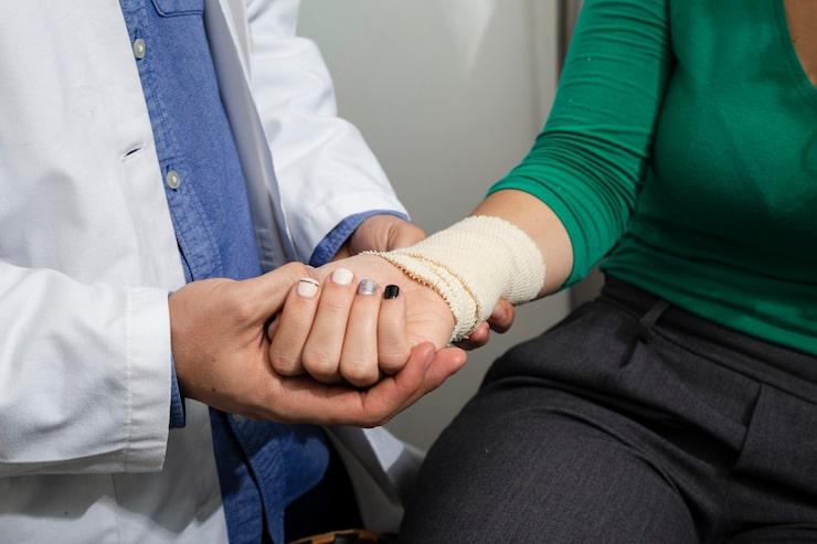
Learn about stress fractures, including causes, symptoms, treatment options, and recovery tips for faster healing and injury prevention.
18 May 2026
by Prakash Hospital

Learn what causes heart palpitations and when they signal heart trouble. Expert insights and treatment from cardiologists at Prakash Hospital.
14 Nov 2025
by Prakash Hospital

High cholesterol increases heart disease risk. Know causes, tests, and how to lower it. Visit Prakash Hospital Noida for cardiology care.
2 Jul 2026
by Prakash Hospital

जानिए लो हीमोग्लोबिन का दिल की सेहत पर क्या प्रभाव पड़ता है। इसके कारण, लक्षण और समय पर इलाज व सही खानपान से कैसे बचाव किया जा सकता है, सरल भाषा में समझें।
6 May 2026
by Prakash Hospital
Answers to common questions about Cardiac Catheterisation (Right and Left Heart Study) at Prakash Hospital, Noida.


Book a consultation with Prakash Hospital's specialists — 24/7 emergency care, 100+ doctors, NABH accredited.
View All Departments