


The Link Between Stress and Heart Disease
Learn symptoms, risk factors, prevention tips and when to seek expert cardiac care in Noida.
25 Mar 2026
by Prakash Hospital
An ultrasound scan of your heart that shows how well it pumps and whether its valves open and close properly, with no needles and no radiation.

Your ankles swell by evening and you're breathless by the first-floor landing. Or you've just had a heart attack, and everyone wants to know how much damage it did. Either way, the next step is very often an echo.
Echocardiography is an ultrasound of the heart, using the same kind of probe as a pregnancy scan. Moved over your chest with some gel, it builds a moving picture of the chambers, walls and valves. Doppler adds the direction and speed of blood flow, which shows leaking or narrowed valves and helps estimate the pressure in your lungs.
An echo shows how strongly the heart pumps, whether the muscle is thickened or damaged, how each valve is working and whether there's fluid around the heart. It can't see inside the coronary arteries, so a normal echo doesn't rule out a blockage.
Our cardiology unit has 2D echocardiography with Doppler. For people with a known heart condition, we repeat the echo at intervals to track changes and adjust treatment.
An echo is used to find the cause of symptoms and to follow known heart problems over time.
The probe stays on the outside of your chest (a transthoracic echo). It answers most questions and is the test most people need.
Pictures are taken before and after exercise, or after a medicine that makes the heart work harder. Muscle that stops moving well under stress suggests a narrowed artery.
A thin probe passes down the food pipe, which lies right behind the heart, after a throat spray and sedation. It gives clearer views of the valves and of clots, and it's arranged only when a standard echo can't answer the question.
You lie on the couch in a gown, usually turned onto your left side, with three ECG stickers on your chest.
Cool gel goes on, and the probe is pressed between the ribs, under the breastbone and sometimes at the base of the neck. Firm pressure can be uncomfortable for a moment but shouldn't hurt.
Any whooshing sounds are the blood flow being measured. You may be asked to hold your breath or roll further over for a clearer view.
The gel is wiped off and you can dress and leave. Most scans take 20 to 40 minutes.
The report describes each chamber and valve and ends with a short impression, which is the part to read first. Your cardiologist usually goes through it with you the same day.
The number most people look for is the ejection fraction (EF), the share of blood the main pumping chamber pushes out with each beat. About 55% or more is usually normal and 50 to 54% is borderline. From 41% to 49% is mildly reduced, and 40% or below means a weak heart muscle. "Regional wall motion abnormality" means part of the wall isn't moving well, often after a heart attack. "LVH" is thickened muscle, commonly from high blood pressure. Grade 1 diastolic dysfunction, a stiffer relaxing phase, is very common after 50.
Valves are graded for leakage (regurgitation) or narrowing (stenosis) as trivial, mild, moderate or severe. Trivial and mild leaks are common in healthy people. Moderate or severe findings usually mean treatment, closer follow-up or further tests such as a coronary angiogram.
After a standard or stress echo you can eat, work and drive as normal.
Wait about an hour before eating or drinking, until your throat is no longer numb. Don't drive or ride for the rest of the day because of the sedation.
If a problem is found, your cardiologist will explain the next step, which may be a change in medicines, another test or a repeat echo in 6 to 12 months.
Call us on +91-8826000033 or come straight to our 24/7 emergency department.
A 10-second electrical tracing. It picks up rhythm problems and old heart attacks but says little about the valves or pumping strength.
Gives very detailed pictures of the heart muscle, which helps when echo images are unclear or when inflammation or scarring is suspected. Some metal implants rule it out.
An echo can't show narrowed coronary arteries. A treadmill test, CT coronary angiogram or catheter angiogram answers that question.
Talk to our Cardiology team
Ask about your options, the cost and your insurance before you decide. See the insurance and TPA panels we work with.
This page explains Echocardiography (2D Echo with Doppler) in general terms. Your own plan depends on your health, your test results and your doctor's advice. Please read our medical disclaimer.
Echocardiography (2D Echo with Doppler) is led by our Cardiology team, with other departments stepping in at each stage of your care.
At Prakash Hospital, our Cardiology Department offers expert, round-the-clock care, providing timely diagnosis and advanced treatments for heart conditions to keep your heart healthy and strong.
What the department treats
Interventional Cardiology
Echocardiography
Cardiac Rehabilitation
Pacemaker Implantation
Angioplasty and Stenting
Heart Failure Management
Procedures often considered alongside Echocardiography (2D Echo with Doppler), or that treat related conditions.
An ECG recorded while you walk on a treadmill, to check for reduced blood flow to the heart during exercise.
A portable ECG worn for 24 to 48 hours to catch irregular heartbeats that come and go.
An X-ray dye test that maps blockages in the heart's arteries and guides the choice between medicines, stents and surgery.
Measures pressure and oxygen inside the heart and lungs through a thin tube, to guide valve and heart failure care.
A small device under the collarbone that keeps a slow or pausing heartbeat at a safe rate.
Other procedures our Cardiology team performs.
We offer expert care across key specialties, including Medicine, Cardiology, Orthopaedics, ENT, Gynaecology, and more—delivering trusted treatment under one roof.
Free tools to check your weight, plan a pregnancy, track calories and more.
Learn symptoms, risk factors, prevention tips and when to seek expert cardiac care in Noida.
25 Mar 2026
by Prakash Hospital

Understand the critical difference between a heart attack and cardiac arrest, their symptoms, causes, emergency response steps, and treatment options. Learn when to seek urgent help from expert cardiologists at Prakash Hospital.
8 Dec 2025
by Prakash Hospital

Understanding early symptoms of heart disease can help prevent severe complications. Learn about the key warning signs such as chest discomfort, fatigue, and breathlessness in this in-depth guide.
23 Jul 2025
by Prakash Hospital

Skin pigmentation — what causes dark patches, melasma, and uneven skin tone, and what treatment works. Prakash Hospital Noida.
9 Sept 2026
by Prakash Hospital

Best Indian foods for healthy, glowing skin — what to eat for skin hydration, collagen, and protection from UV damage. Prakash Hospital Noida.
14 Sept 2026
by Prakash Hospital

Oily skin causes in India, what makes it worse, and how to manage it with diet and skincare. Prakash Hospital Noida.
11 Sept 2026
by Prakash Hospital
Learn what causes heart palpitations and when they signal heart trouble. Expert insights and treatment from cardiologists at Prakash Hospital.
14 Nov 2025
by Prakash Hospital
दिल को लंबे समय तक स्वस्थ रखने के लिए जानिए 10 आसान और असरदार टिप्स। हार्ट डिज़ीज़ से बचाव, सही डाइट और लाइफस्टाइल गाइड
24 Dec 2025
by Prakash Hospital
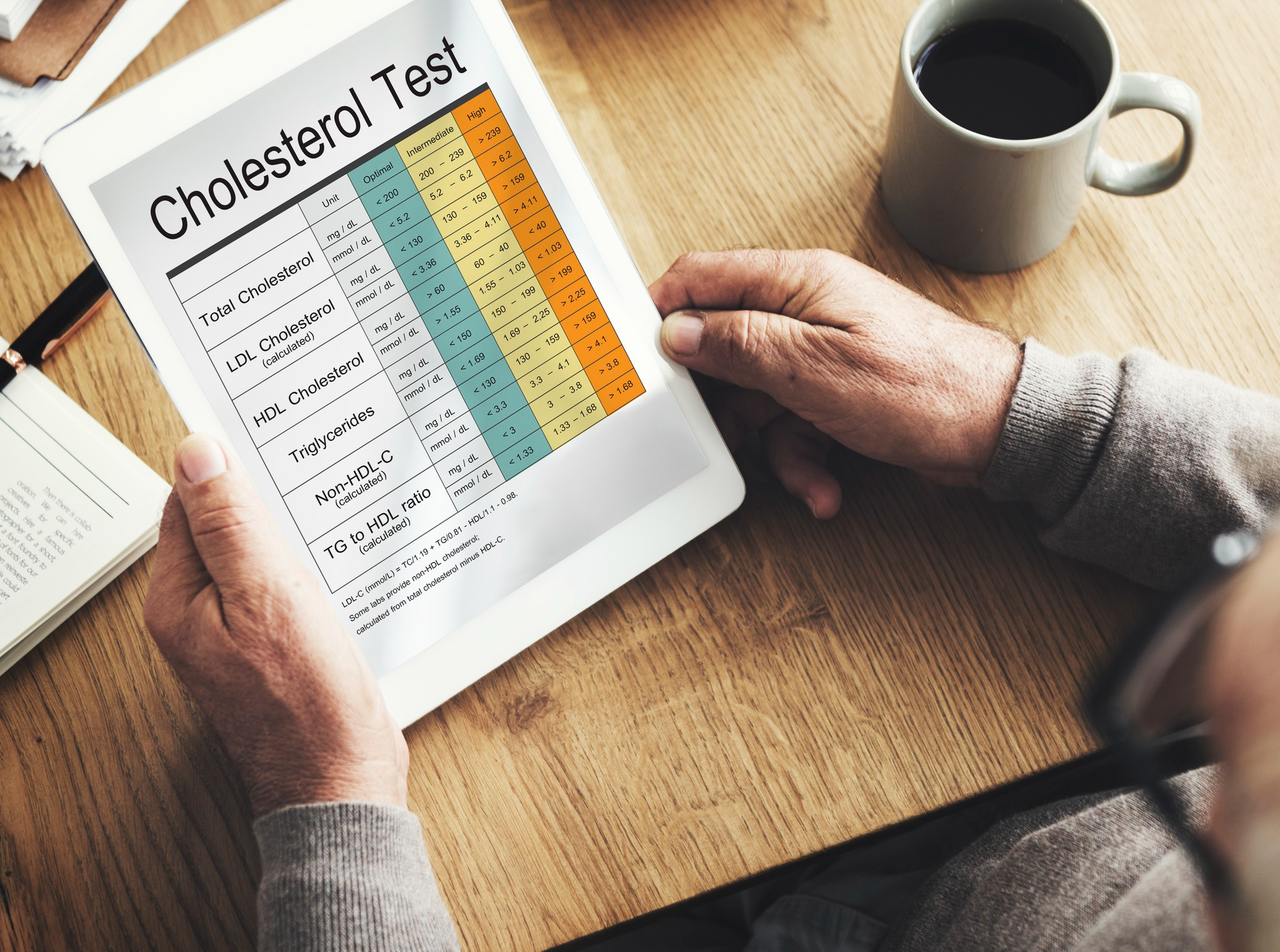
Understand HDL and LDL cholesterol, why balance matters, and how to support heart health effectively.
1 Dec 2025
by Prakash Hospital
Answers to common questions about Echocardiography (2D Echo with Doppler) at Prakash Hospital, Noida.


Book a consultation with Prakash Hospital's specialists — 24/7 emergency care, 100+ doctors, NABH accredited.
View All Departments